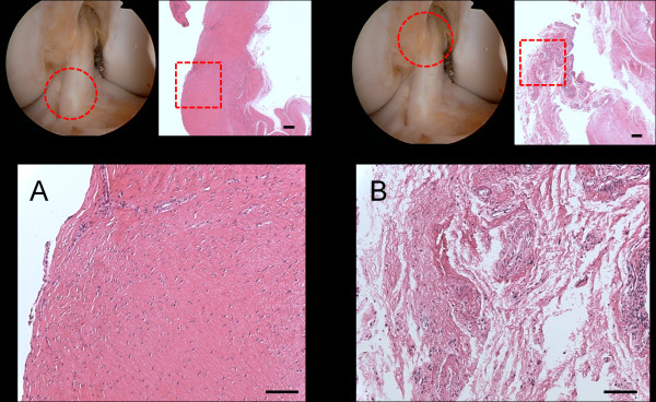
Figure 3.
Photomicrographs of sections of the extracted graft. (Hematoxyline and eosin stains). (A) The distal non-impinged part of the ACL graft showing normal synovial coverage and regular pattern of the collagen fibers. (Superior left: arthroscopic view, Superior right: original magnification × 25, Scale bar = 200 μm, Inferior: original magnification × 100, Scale bar = 100 μm) (B) The proximal impinged part against PCL showing enhanced vascularization and proliferation of the overlying synovium. The sections showed irregularity of the collagen fibers and scattered hyaline degeneration of the graft. (Superior left: arthroscopic view, Superior right: original magnification × 25, Scale bar = 200 μm, Inferior: original magnification × 100, Scale bar = 100 μm).

