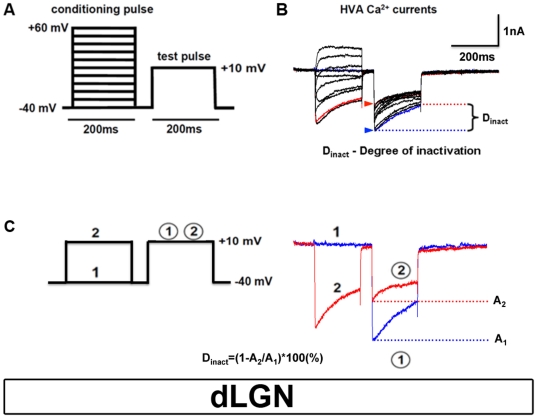
Figure 3. Identification of CDI in TC neurons.
(A) Scheme of the double-pulse protocol used to elicit HVA Ca2+ currents. TC neurons were held at −40 mV and conditioning pulses to varying potentials (−40 to +60 mV, 200 ms duration) were followed by a brief gap (−40 mV, 50 ms) and a subsequent analyzing test pulse to a fixed potential of +10 mV (200 ms). (B) Family of representative current traces elicited by the pulse protocol shown in (A). (C) Dinact equation for calculating effects of CDI in TC neurons.

