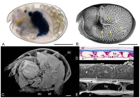
Figure 6. General morphology of Recent ostracods exemplified by myodocopids.
A, B, Vargula hilgendorfii (Müller), left lateral view of live female carrying embryos in her domiciliar cavity (carapace translucent) and lateral view of left valve in transmitted light showing the gas-exchange area, an integumental hemolymph network (yellow arrows indicate hemolymph circulation). C, Azygocypridina sp. from New Caledonia, France. Scanning electron micrograph showing appendages, left valve removed, including the ventilatory plates (courtesy of Vincent Perrier). D, E, transverse section through the carapace of Vargula hilgendorfii showing hemolymph sinuses, stained microtome serial section and scanning electron micrograph, respectively (see [51], [76]). Scale bars: 1 mm in A–C, and 20 µm in D and E. ef, epipodial fan for ventilation; hs, hemolymph sinus; il, inner lamella; ol, outer lamella.

