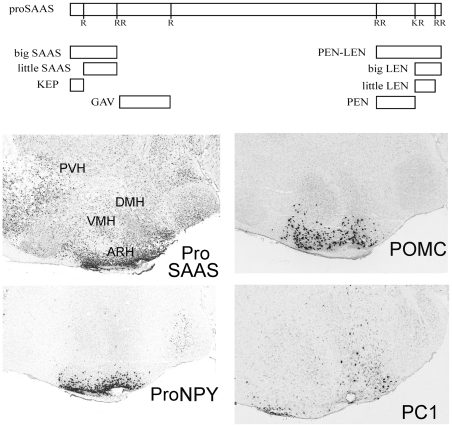
Figure 1. Schematic of proSAAS and proSAAS-derived peptides, and the localization of proSAAS, proNPY, POMC, and PC1/3 mRNA in the hypothalamus of the mouse.
Top panel: Schematic diagram showing the major proSAAS-derived peptides previously detected in various brain regions and other relevant peptides. The relative size and position of these peptides within proSAAS are indicated. Bottom panels: Images of proSAAS, proNPY, POMC, and PC1/3 mRNA distribution were downloaded from the Allen Mouse Brain Atlas [http://mouse.brain-map.org], Seattle, WA, Allen Institute for Brain Science ©2009 [50].

