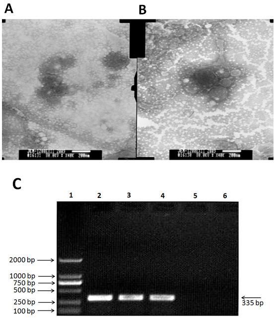
Figure 1.

Identification of ZJ7 isolate by morphology and molecular biology methods. (A and B) The morphology of ZJ7 isolate under electron microscope (negative staining) and the particles of assembling viruses; (C) Electrophorogram results of RT-PCR from 3 and 6 passages, the band is 335bp, that is same size as the fragment we designed.
