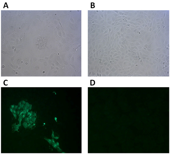
Figure 2.

Microphotographs and IFA results. (A) CPE in MDCK cell with large round syncytium and multiple nuclei by CDV-ZJ7. (B) Typical CDV destruction of cell layers for mock. (C) IFA for ZJ7 isolate-infected MDCK cell and (D) for Non-infected MDCK cells. Original magnifications is 200 ×.
