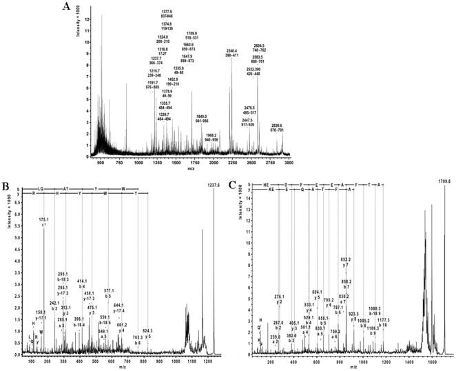
Figure 3. Identification of 60 kDa band.
A. The PMF spectra of tryptic fragments of 60 kDa glycoprotein. PMF spectra of tryptic fragments of 60 kDa were identified as N-terminal fragment of α-spectrin by MALDI-TOF MS. Each fragment is denoted by their m/z values and sequence range within the 955 amino acids of human α-spectrin (marked with yellow in Fig. S1). B–C. Confirmation of the sequence of the identified tryptic fragments by MALDI-TOF-TOF mass spectrometry. The MS/MS spectrum was analyzed with database-dependent MASCOT as well as database-independent Sequit! software systems yielding the same results. Two representative PSD spectra of the MS/MS analysis of the fragment (B) LQATYWYHR (m/z = 1237.6) and (C) HEDFEEAFTAQEEK (m/z = 1237.6) of α-spectrin and SGP-60. The N and C terminal fragment ions are denoted according to standard nomenclature and immonium ions displayed in single amino acid code.

