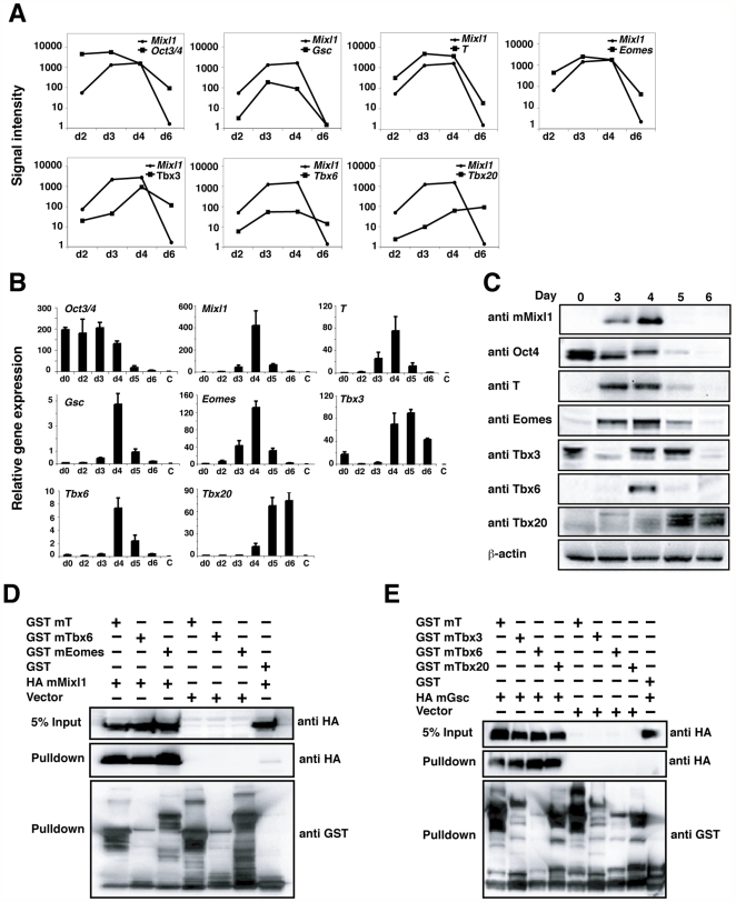
Figure 4. Tbx factors are co-expressed with Mixl1 during ESC differentiation and interact with Mixl1.
(A) Signal intensity of Mixl1, Oct3/4, Gsc and T and the Tbx genes Eomes, Tbx3, Tbx6 and Tbx20 during ESC differentiation, as measured by microarray analysis. Expression profiles of the genes in (A) were validated by real time-PCR (B) results from an independent experiment are shown. Error bars show the S.E.M., n = 3 and Western blot analysis (C). (D) Mixl1 interacts with members of the Tbx family. 293T cells were transfected with HA mMixl1 and GST mT, mTbx6 or mEomes expression plasmids as indicated. GST-fusion proteins were isolated from whole cell extracts using glutathione resin and bound fractions were analysed by Western blot with anti-HA antibody. Expression of each protein was confirmed with anti-GST and anti-HA antibodies. (E) Gsc interacts with members of the Tbx family. 293T cells were transfected with HA mGsc and GST mT, mTbx3, mTbx6 or mTbx20 expression plasmids. Reactions were performed as in (D).

