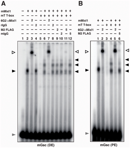
Figure 5. Interaction between T and DNA bound Mixl1.
(A) Mixl1 and T form a complex on the Gsc promoter distal element (DE). EMSA was performed with Gsc promoter DE probe and 50 ng His-tagged Mixl1 protein (lanes 2 to 4 and 6 to 12). Samples in lanes 5 to 12 included 200 ng FLAG-His-tagged T T-box domain protein. The presence of Mixl1 and T in the DNA complexes was confirmed by super shift reactions using antibodies against Mixl1 (lanes 3 and 7) or the FLAG epitope (lanes 9 and 11). Lanes 3 and 7 received 2 µg 6G2 anti-Mixl1 antibody and lanes 4 and 8 received 2 µg rat isotype IgG. Lanes 9 and 11 received 2 µg and 5 µg of M2 FLAG antibody, showing that 5 µg of M2 FLAG antibody was required to demonstrate supershift activity. Lanes 10 and 12 received 2 µg and 5 µg of mouse IgG antibody, respectively. (B) Mixl1 and T form a complex on the Gsc promoter proximal element (PE). EMSA was performed as above except the Gsc PE MBS probe was used. Samples represented by lanes 1, 2 and 4 to 6 included 50 ng His-tagged Mixl1 protein whilst those present in lanes 3 to 6 contained 200 ng FLAG-His-tagged T T-box domain protein. Super shift reactions were performed by adding 6G2 anti-Mixl1 antibody (2 µg) to samples in lanes 2 and 5 or M2 FLAG antibody (5 µg) to the sample in lane 6. The black arrowheads indicate the position of the Mixl1 and Mixl1-T-DNA complexes; the white arrowhead shows the complexes super-shifted in the presence of the anti-6G2 or M2 FLAG antibodies; the grey arrowhead indicates the free probe.

