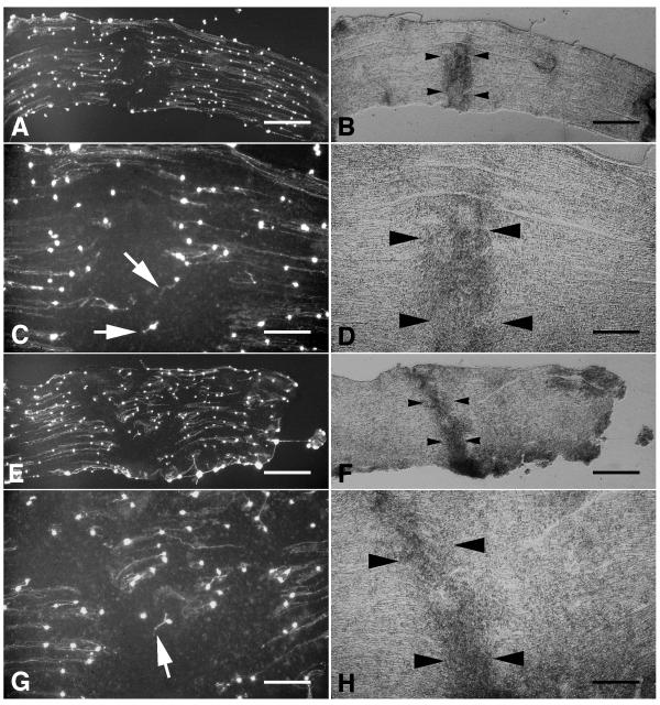
Figure 4.
Neurite growth on ex vivo crushed sciatic nerve. A,E, Fluorescein-labelled neurites extending on longitudinal sections through crushed sciatic nerves. Long parallel neurites extended on uncrushed portions of the nerve sections but did not extend onto the crushed segments. Higher power photomicrographs of the crushed segments of the sections in panels A and E are shown in panels C and G, respectively. Neurites extending from neurons attached directly on crushed nerve tissue were usually shorter and oriented in non-parallel orientations (white arrows). B, D,FH,, Phase-contrast photomicrographs of the same fields shown in panels AC,,EG,, respectively. Crushed sciatic nerve is evident from its increased optical density. The edges of the crushed segments of sciatic nerve are indicated by black arrowheads. Scale bars: A, B, 500 μm; C, D, 200 μm; E, F, 500 μm; G, H, 200 μm.

