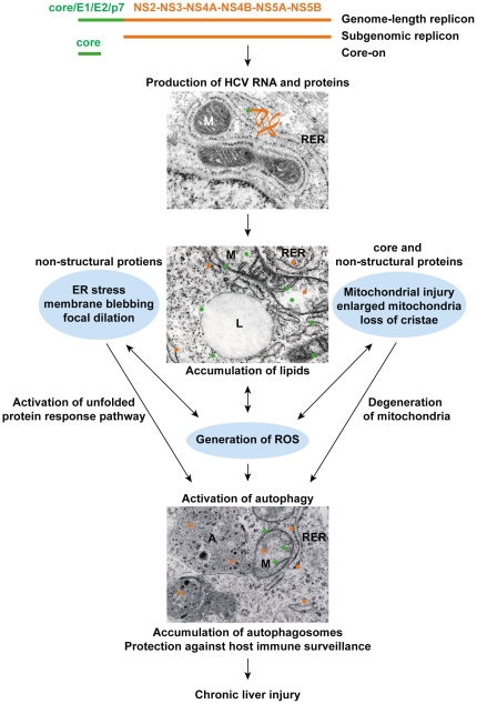
Figure 7. Diagrammatic presentation of the cytopathic effects caused by various HCV proteins.
Replication of HCV RNA and production of HCV structural (core, E1, E2, and p7) and non-structural proteins (NS2, NS3, NS4A, NS4B, NS5A, and NS5B) in the rough ER and the subsequent partition of HCV proteins to different subcellular compartments lead to ER stress, mitochondrial injury, and the production of ROS. These cytopathic effects lead to activation of autophagy without a concomitant increase in protein degradation. Consequently, autophagosomes accumulate in HCV infected cells. EM photo with normal mitochondria (M) and rough ER (RER) was taken from healthy Huh7 cells; EM photos with enlarged mitochondria, ER blebbing, lipid droplet (L), and autophagosomes (A) were taken from genome-length and subgenomic replicon cells. The HCV genetic materials contained in genome-length replicon, subgenomic replicon, and Core-on cells are depicted at the top of the diagram.

