
Figure 3.
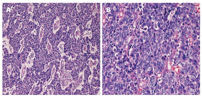
Polypoid tumor. A: The polypoid tumor consists of malignant cells with hyperchromatic nuclei arranged in a trabecular pattern (HE, × 100); B: Higher power view shows large cells, vesicular nuclei, nucleoli, mitotic figures and apoptotic bodies (HE, × 200). HE: Hematoxylin and eosin.