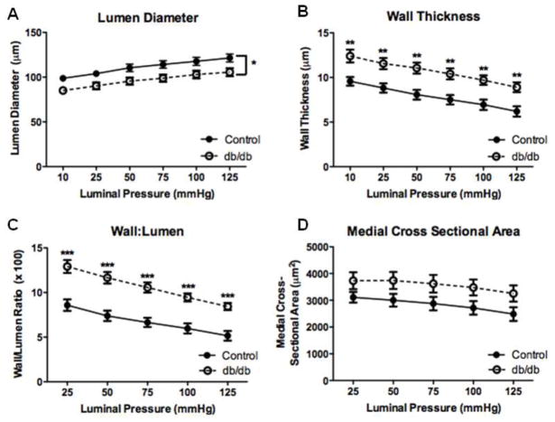
Fig. 2.
Passive structural measurements of isolated coronary arterioles from 16-wk diabetic and control mice. These measurements demonstrated decreased lumen diameter (a), increased wall thickness (b), increased wall-to-lumen ratio (c) and no change in medial CSA (d). Values are mean SEM, * p < 0.05, ** p < 0.01, and *** p < 0.001vs. control.

