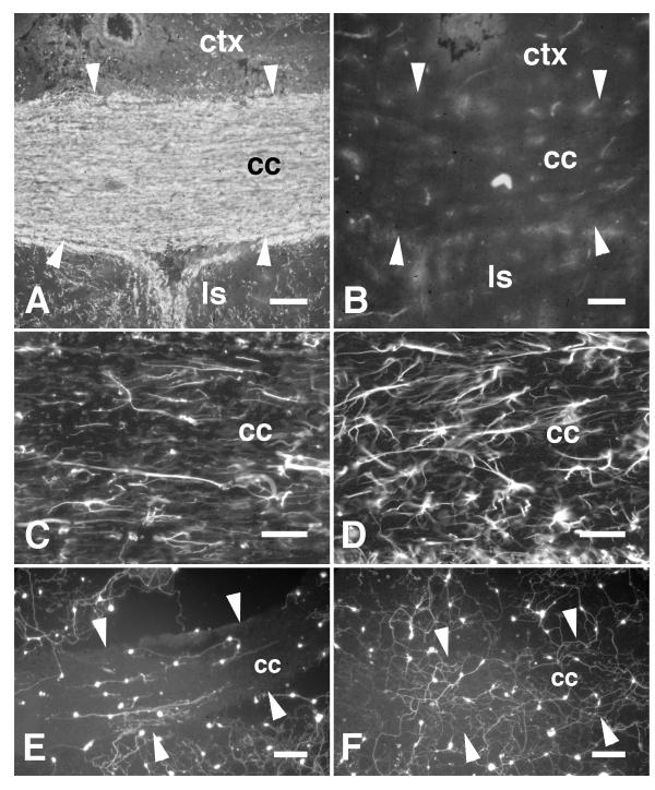
Figure 3.
Neurites extending on myelin-deficient corpus callosum. A, The corpus callosum (cc) of a wild-type rat (21 days old) is shown labelled by the myelin-specific antibody Rip. The contrast in myelin content between the corpus callosum and gray matter such as the neocortex (ctx) and lateral septal nuclei (ls) is evident. B, The corpus callosum (cc; edges of the tract are indicated by white arrowheads) of an age-matched myelin-deficient mutant rat labelled with the Rip antibody. In comparison with the wild-type animal, little Rip-immunoreactivity could be detected in the corpus callosum and there was little contrast between gray and white matter indicating the absence of myelin. C, An adjacent section showing the corpus callosum of the wild-type rat labelled by GFAP-immunohistochemistry showing mostly parallel orientations to the astroglial processes. D, An adjacent section showing the corpus callosum of the myelin-deficient rat labelled by GFAP-immunohistochemistry. The astrocytes appeared hypertrophic but are mostly oriented in parallel with the fiber tract. E, Fluorescein-labelled neurons cultured on a section through the wild-type corpus callosum. Neurites extending on white matter were oriented in parallel with the tract. Neurites extending on gray matter rarely extended across borders with white matter. F, Neurons cultured on a section through the myelin-deficient corpus callosum. The neurites were unoriented on white matter and frequently extended across borders between white and gray matter. Scale bars: A, B, 100 μm; C, D, 50 μm; E, F, 150 μm.

