
Fig 1.
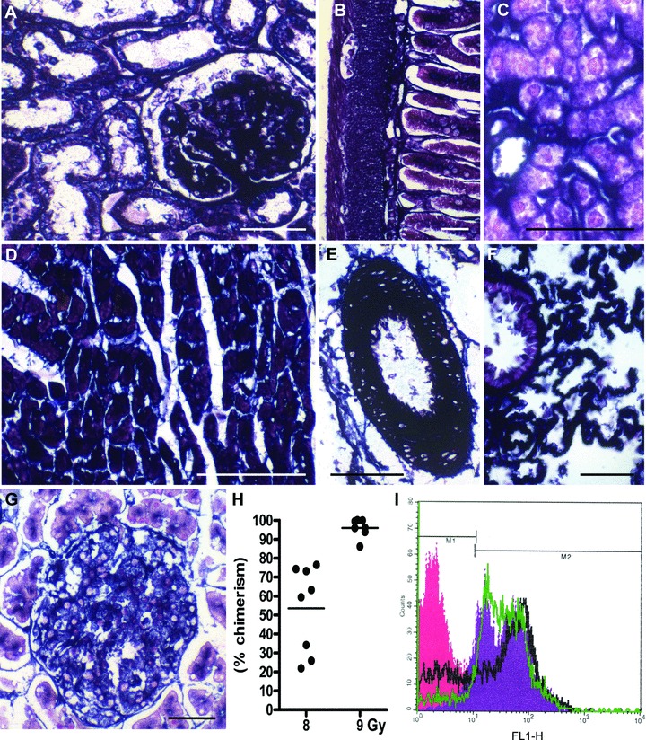
Expression of the marker enzyme ALPP in tissues of ALPP-transgenic (ALPP-tg) F344 rats and chimerism after reconstitution of irradiated wt rats with ALPP-tg BM. (A)–(G) Histochemical staining showed strong and ubiquitous expression of ALPP in cells of epithelial, endothelial and mesenchymal origin in kidney (A), gut (B), liver (C), heart muscle (D), arteries (E), lung (F), as well as exocrine and endocrine pancreas (G) in ALPP-tg rats. The 5-μm-thick paraffin sections shown in (A)–(G) were stained for ALPP enzyme activity (BCIP/NBT, purple) overnight at RT after heat pre-treatment, and were counterstained with nuclear fast red. Bars represent 50 μm. (H)–(I) FACS analysis of BMC isolated from wt rats lethally irradiated with a linear accelerator, using a single dose of 8 or 9 Gy, and reconstituted with unfractionated BM harvested from ALPP-tg donors. Irradiation with 9 Gy resulted in high ALPP chimerism (96.9 ± 6.4%), whereas chimerism was much lower using a single dose of 8 Gy (53.6 ± 22.7%). Chimerism was determined on the basis of a standard curve. Representative single parameter fluorescence histogram (I) shows hPLAP expression (marker M2) on nearly all BMC in reconstituted wt rats after irradiation with 9 Gy (green line), similar to BMC from hPLAP-tg rats (violet area). Irradiation with 8 Gy (black line) resulted in lower degree of chimerism. The red area depicts ALPP– (marker M1) BMC from wt control rats.