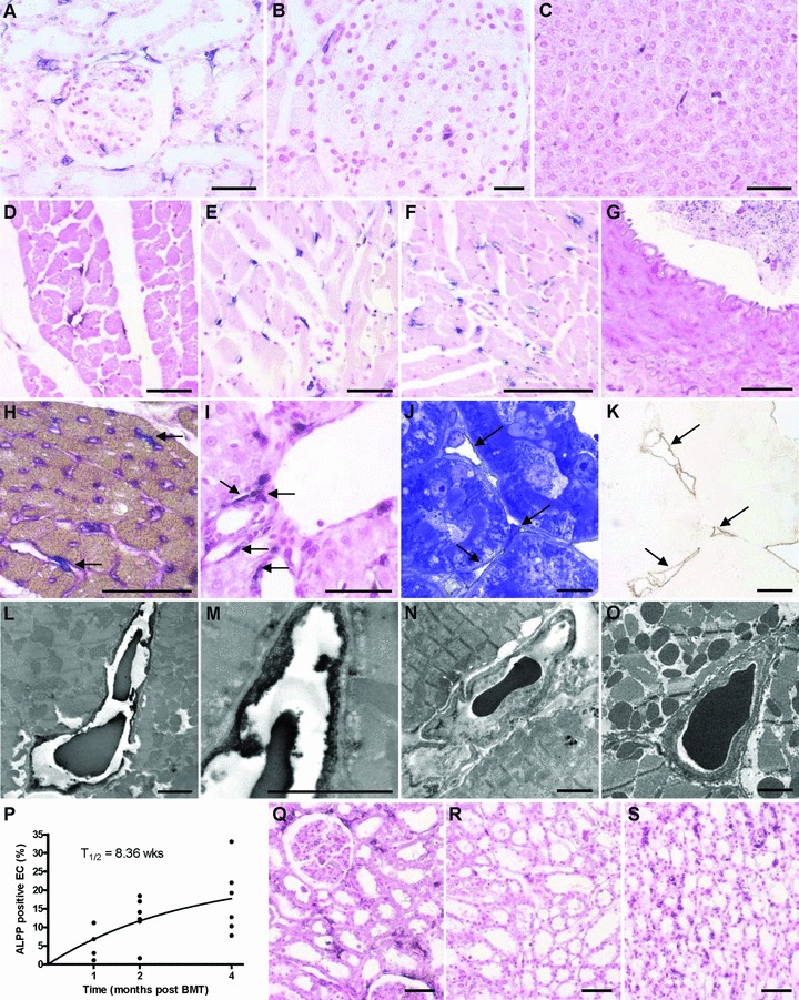
Fig 2.

Light and electron microscopic analysis of tissues from wt F344 rats, lethally irradiated and reconstituted with BM from ALPP-tg F344 donors. ALPP+ endothelial-like cells in capillaries are evident in kidney (A), pancreas (B) and liver (C), 2 months after BM transplantation (BMT). The number of ALPP+ endothelial-like cells increased with time in all organs as shown for heart tissue 1 (D), 2 (E) and 4 (F) months after BMT. In contrast, ALPP+ EC were absent in large blood vessels such as the Aorta thoracica (G), arteries or veins (not shown). (H) Double staining (arrows) for ALPP by histochemistry (blue) and for tomato lectin Lycopersicon esculentum (purple) confirmed the endothelial nature of the putative BM-derived capillary EC in the myocardium, 2 months after BMT. (I) ALPP+ cells in the vicinity of blood vessels as shown here in liver. Semi-thin serial sections of epon-embedded, 45-μm-thick kidney cryosections stained immunohistochemically against ALPP by a pre-embedding protocol and counterstained with toluidine blue (J) or left without counterstaining (K, brown DAB immunoprecipitate) show that the anti-ALPP staining (arrows in K) co-localized with capillaries situated between renal tubuli (arrows in J), 6 months after BMT. (L)–(O) Transmission electron microscopy (TEM) of ultra-thin sections of the heart muscle after pre-embedding anti-ALPP staining clearly showed that the BM-derived ALPP+ endothelial-like cells are indeed capillary EC, 6 months after BMT. Higher magnification is shown in (M). Anti-ALPP staining was absent in control sections of hearts from BMT rats when the primary antibody was omitted (N) or in wt controls (O). (P) Nonlinear regression analysis of the time dependent increase in BM-derived EC in the heart muscle revealed a half time of 8.36 weeks for these cells. (Q)–(S) In the kidney, EC turnover showed marked inhomogeneity. BM-derived ALPP+ EC were more frequent in cortical regions (Q) and in the Vasa recta of the papilla (S) than in medullary areas (R) throughout the study period, as demonstrated here at 2 months after BMT. The 5-μm-thick paraffin sections shown in (A)–(G), (I) and (Q)–(S) were stained for ALPP enzyme activity with BCIP/NBT (purple) overnight at RT after heat pre-treatment, and were counterstained with nuclear fast red. Semi-thin and ultra-thin sections shown in (J)–(O) were immunostained using a monoclonal anti-ALPP antibody by the pre-embedding protocol described in ‘Methods’. Bars represent 50 μm for light microscopy and 2.5 μm for transmission electron microscopy.
