
Fig. 1.
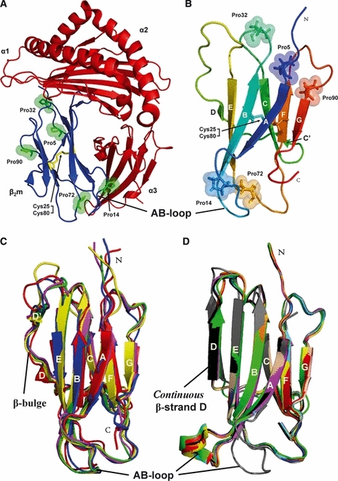
Monomeric β2m plays a key role in DRA. (A) Cartoon representation of human MHC I (PDB code 3MYJ [136]) showing the heavy chain (α1, α2, α3 in red) and the light chain (β2m in blue). Highlighted are the residues Pro5, Pro14, Pro32, Pro72 and Pro90 (in green sticks, spheres) and the disulfide bond between residues Cys25 and Cys80 (in yellow sticks). (B) Cartoon representation of the solution structure of monomeric native wild-type β2m (PDB code 2XKS [9]) showing β-strands A (6–11), B (21–28), C (36–41), C′ (44–45), D (50–51), E (64–70), F (79–83) and G (91–94). Highlighted are the residues Pro5, Pro14, Pro32, Pro72 and Pro90 (in sticks, spheres) and the disulfide bond between residues Cys25 and Cys80 (in sticks). N, N-terminus; C, C-terminus. (C) Structures displaying a β-bulge and an attached AB-loop: wild-type β2m (PDB code 1JNJ [7]) in red, H31Y (PDB code 1PY4 [15]) in green, W60G (PDB code 2VB5 [16]) in blue, H13F (PDB code 3CIQ [55]) in yellow and MHC I (PDB code 3MYJ [136]) in magenta. (D) Structures displaying a straight β-strand D: wild-type β2m (PDB code 1LDS [11]) in red, L39W/W60F/W95F (PDB code 2D4D [137]) in green, wild-type β2m (PDB code 2D4F [137]) in blue, wild-type β2m (PDB code 2YXF [12]) in yellow, W60G (PDB code 2Z9T [16]) in magenta, W60C (PDB code 3DHJ [14]) in cyan, D59P (PDB code 3DHM [14]) in orange, W60G (PDB code 3EKC [14]) in wheat, K58P/W60G (PDB code 3IB4 [121]) in black and P32A (PDB code 2F8O [58]) in grey.