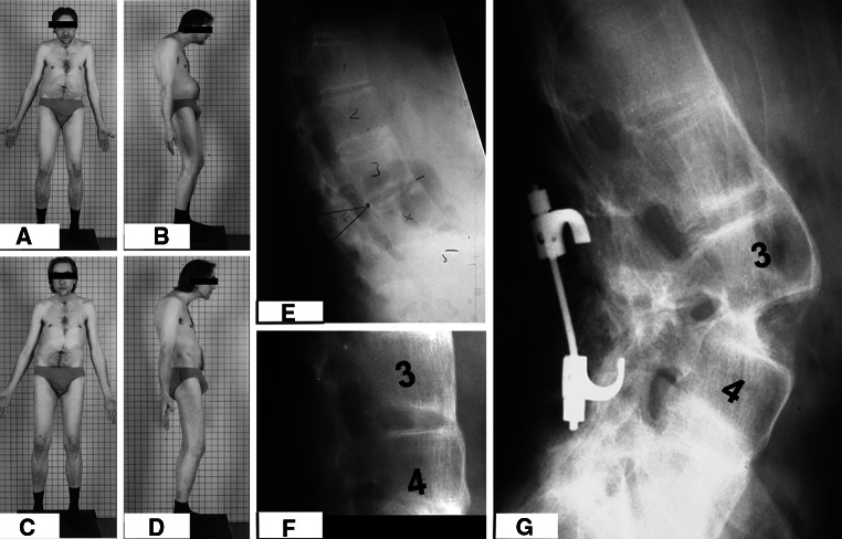
Fig. 1.
a, b The pre-operative photographs of a patient with AS and TKLD. Notice the protuberance of the abdomen on the lateral view. c, d The post-operative photographs following OWO technique. The patient worked as a cabinet maker and hence maximal correction was not performed to ensure that the patient had adequate horizontal gaze and also a functional degree of inferior vision to aid his profession. e The pre-operative radiographs of the same patient with TKLD, the opening wedge was planned at L3–4 interspace (f). f The follow-up radiograph with healed osteotomy site, correction was stabilized with hook and rod system

