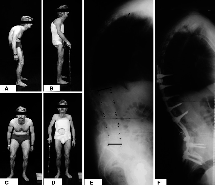
Fig. 2.
a, b The lateral pre- and post-operative photographs, respectively of an AS patient with TKLD, who underwent correction using the CWO technique. c, d The frontal photographs in the same patient pre- and post-op, notice the compressive effect on the abdomen by the inferior margins of the ribs which has been relieved following surgery. e The pre-operative lateral radiographs in the same patient, notice the TKLD and the loss of lumbar lordosis. f The post-operative lateral radiographs in the same patient, who underwent corrective osteotomy using CWO technique and instrumentation using pedicle screws and solid contoured rods

