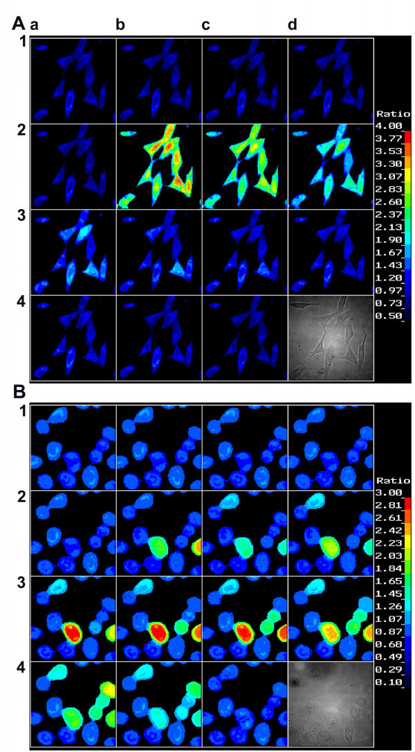
Figure 1.
Effect of ethylene on [Ca2+]i level in NIH-3T3 cells (A) and SaOS-2 cells (B). Cells were loaded with Fura-2 and analyzed by the fluorescence ratio-imaging system as described in Materials and Methods. Ethylene was generated by addition of 1 mM ethephon (time: 5.0 min). Fluorescence images were recorded at time zero (a1) and after 1.5 (b1), 3 (c1), 4.5 (d1), 5 (a2), 5.5 (b2), 6 (c2), 6.5 (d2), 7 (a3), 7.5 (b3), 8 (c3), 8.5 (d3), 10 (a4), 11.5 (b4), and 13.5 min (c4). In (d4) the cells inspected are shown by Nomarski phase contrast interference optics. The spectrum color scale ranges from blue (low [Ca2+]i) to red (high [Ca2+]i). Magnification, 400-fold.

