Figure 1.
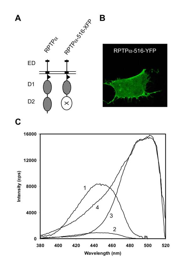
Expression and spectral properties of chimeric fluorescent RPTPα proteins. (A) Schematic representation of wild type RPTPα (RPTPα and RPTPα-516-XFP, a construct in which either CFP or YFP (indicated by XFP) was inserted at position 516, thereby replacing the membrane distal PTP domain, D2. The extracellular domain (ED), transmembrane domain (black box), membrane-proximal domain (D1) and membrane-distal domain (D2) are indicated. (B) Confocal micrograph of a single SK-N-MC neuroepithelioma cell transfected with an expression vector for RPTPα-516-YFP, illustrating membrane localization of RPTPα-516-YFP. (C) SK-N-MC cells were transfected with RPTPα-516-CFP (traces 1 and 2), RPTPα-516-YFP (trace 3), or both (trace 4). Excitation spectra of single, live cells were corrected for background and are depicted using filter #1 (longpass 490 nm)(trace 1), allowing detection of CFP, and using filter #2 (bandpass 530-560 nm)(traces 2 - 4), optimal for detection of YFP. Filter #2 is the FRET filter, allowing detection of FRET when CFP and YFP are in a protein complex.

