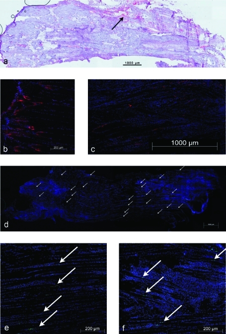
Figure 7.
a. Factor VIII staining of a longitudinally sectioned soleus muscle 4 weeks after crush injury, showing the distribution of vessels in the injury zones. b. Cryo-section with an immunohistochemical stain for nestin in a healthy soleus muscle (counterstain: DAPI), myotendinous transition zone. c. Staining for nestin in a longitudinally sectioned soleus muscle (counterstain: DAPI). Nestin immunoreactivity was dispersed throughout the muscle and could be found not only at the tips of regenerating myofiber stumps but also at the lateral aspects of the fibers. d. Overview of a-bungarotoxin staining of a longitudinally sectioned soleus muscle (counterstain: DAPI). The white arrows indicate multiple, newly developed neural endplates in the proximal and distal crush zones of the muscle. e. Detail of panel d depicting a linear distribution of endplates in the uninjured region. f. Detail of panel d depicting endplates within the crushed area.

