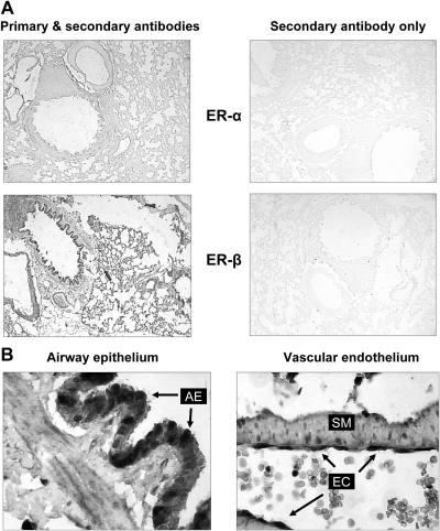
Fig. 2.
Immunohistochemical localization of ER in rat lungs. A, low-magnification view (×40) of immunohistochemical staining for ERα and ERβ in male rat lung. Sections were incubated in the presence (left panel) or absence (right panel) of primary antibody to demonstrate specificity of staining. B, Strong immunohistochemical staining of ERα is noted in airway epithelial cells (AE, arrows, left panel), and endothelial cells (EC, arrows, right panel), and smooth muscle cells (SM). Original magnification, ×400.

