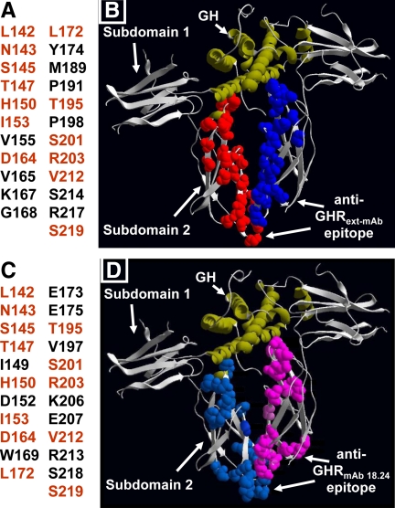
Fig. 6.
Anti-GHRext-mAb and anti-GHRmAb 18.24 epitopes. A and C, Residues in subdomain 2 that when mutated prevent recognition of rabbit GHR by anti-GHRext-mAb (A) and anti-GHRmAb 18.24 (C). Shared residues in both epitopes are shown in orange. B and D, Side chains of residues identified in A and C are shown as space-filling balls (red or light blue for one receptor, blue or pink for the second receptor in the dimer) in the context of the crystal structure of the GH-engaged GHR dimer. GH ribbon structure is shown in green.

