Figure 2.
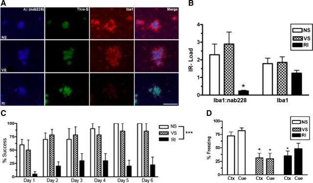
Chronic stress impairs spatial and fear-related learning but does not correlate with activated microglia-associated Aβ plaques in Tg2576 mice. Tg2576 mice were assessed for activated microglia associated with Aβ plaques. A, Representative images show that, regardless of stress condition, all mice display Nab228-IR Aβ plaques (blue) that are Thioflavin-S+ (green) in the frontal cortex. However, RI mice showed significantly less Iba1-IR activated microglia (red) around Aβ plaques without a decrease in overall Iba1-IR. B, This effect is quantified by nonfluorescent immunoreactivity load ratios (mean ± SEM) of adjacent sections (F(2,21) = 6.51, p = 0.007; and F(2,21) = 6.51, p = 0.007; n = 8 per group). Fourteen-month-old Tg2576 mice (n = 10 per group) were also tested on the Barnes maze (C) and fear conditioning (D). RI but not VS mice demonstrated significantly worse percentage success (F(3,35) = 12.8, p < 0.0001) compared with NS mice. In both context (Ctx) (F(2,20) = 6.22, p = 0.009) and cued (Cue) (F(2,21) = 5.98, p = 0.01) fear conditioning, both VS and RI stressed Tg2576 mice froze significantly less than NS mice. Data show mean ± SEM. *p < 0.05, ***p < 0.001 from NS. Scale bar, 100 μm.

