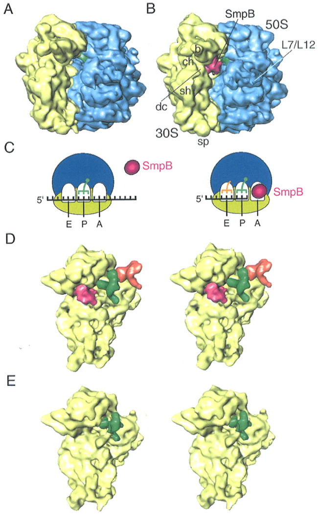
Figure 1.

Cryo-EM maps of SmpB-ribosome complexes.
(A) Cryo-EM map obtained for 70S-tRNA ribosome complex in the presence of an mRNA bearing a sense codon at the A site (sec Experimental Procedures). No density attributable to SmpB appears in the map even though SmpB is in the solution. (B) Cryo-EM map obtained for 70S-tRNA ribosome stalled with a truncated mRNA in the presence of SmpB (see Experimental Procedures). The density attributable to SmpB is shown in red, while the 50S subunit is shown in blue, the 30S subunit in yellow, and the P-site tRNA in green. Landmarks on the 50S subunit: L7/L12, stalk formed by proteins L7/L12. Landmarks on 30S subunit: sh, shoulder; b, beak; dc, decoding center; ch, entrance of mRNA channel; and sp, spur. (C) Schematic recapitulation of the results shown in (A) and (B): SmpB is excluded when a sense codon is in the A site (left panel), while absence of any codon “invites” SmpB to occupy this site (right panel). (D) Stereo view of map computationally separated from the map shown in (B). The 30S subunit is shown in yellow, SmpB in pink, P-site tRNA in green, and E-site tRNA in orange. (E) Stereo view of the 30S subunit computationally separated map from (A). Only the P site is occupied.
