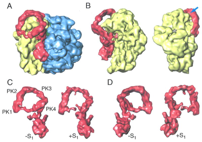
Figure 6.

Cryo-EM map showing the binding between the ribosome and tmRNA in the absence of S1. (A) Cryo-EM map for the EF-Tu·tmRNA·SmpBs complex bound to the ribosome in the absence of S1. The density attributable to the EF-Tu·tmRNA·SmpBs complex is in red. 50S subunit is in blue, the 30S subunit is in yellow. (B) Two views of the 30S subunit bound with EF-Tu·tmRNA·SmpBs in the absence of S1. The arrow points to the contact between PK2 and the head of the 30S subunit. Asterisk: position where ribosomal protein S1 would appear in the 30S subunit. (C) Side view of the tmRNA·SmpBs·EF-Tu·GDP complexes in the absence and presence of S1. (D) View of complexes in (c), after rotation by 180°.
