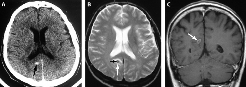
Fig 6 Pathology in neuroschistosomiasis. (A) Unenhanced axial CT scan shows small, oval, hyperdense lesion (black arrow) in the paraventricular zone, dorsal of the right posterior horn. (B) Axial T2-weighted (2437/90/1 [repetition time/echo time/excitations]) MRI shows hypointense lesion (white arrow) with small centrally located area of intermediate signal (black arrow). (C) Coronal contrast-enhanced T1-weighted (600/15/2) MRI shows oval lesion of intermediate signal (arrow) with ring-like and septum-like contrast enhancement. Reprinted from McManus et al13with permission from the American Society for Microbiology. Courtesy of the Wellcome Trust.
