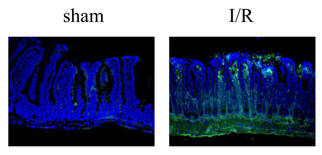
Figure 2.
Deposition of complement C4 in the intestine of I/R treated RAG-1−/− mice reconstituted with human IgM. Representative cryosections were stained with antibody specific for mouse C4 (Green). The left panel is sham treated mouse, and the right panel is I/R treated mouse (400× magnification). All sections were counterstained with DAPI (Violet).

