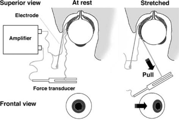
Fig. 1.
Experimental setup: a soft, multi-stranded wire electrode was positioned at the frontal attachment of the lateral rectus muscle and sutured to its fascia. Another wire (100 μm diameter, 1 mm tip without insulation, beveled at ~45°) was gently guided along the wall of the orbit and inserted into the belly of the medial rectus muscle. A polypropylene wire (7.0; 0.5 metric; Prolene, Ethicon) was tied between the frontal muscle attachment and a force transducer. The transducer was pulled manually to rotate the eyeball back and forth in the medio-lateral direction, thus producing cycles of muscle lengthening and shortening, while the force and EMG activity were recorded

