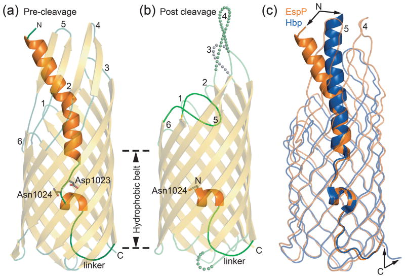
Fig. 1. EspP and Hbp structures.
(a) Pre-cleavage structure of the EspP N1023D mutant. The side chains of Asp1023 and Asn1024 are shown. (b) Post cleavage structure of EspP. The N-terminal residue, Asn1024, is shown. Disordered loops are depicted as spheres. (a and b) β-strands, yellow; α-helices, orange; loops, green. The location of the hydrophobic belt, N and C-termini, extracellular loops 1 through 6, and linker loops are labeled. (c) Structural alignment of pre-cleavage EspP (orange) and pre-cleavage Hbp (blue). For EspP, loops 4 and 5 are labeled. These loops are disordered in the Hbp structure. Figures 1, 2, 5, 6, and 7 were created using The PyMOL Molecular Graphics System (Schrödinger, LLC).

