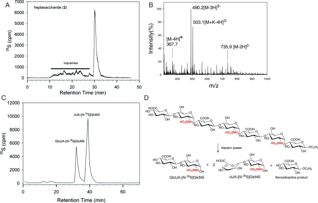
Fig. 4.
Structural characterization of heptasaccharide 2. (A) The HPLC chromatogram of 35S-labeled heptasaccharide 2 using a PAMN column. Solid bar indicates the impurities. (B) The MS spectrum of HPLC-purified heptasaccharide 2. (C) The HPLC of the disaccharide analysis of heparin lyase-digested 35S-labeled heptasaccharide 2. The disaccharide analysis was carried out on a C18-column eluted under RPIP-HPLC conditions. The elution positions of the disaccharides were confirmed with appropriate disaccharide standards. (D) The reactions involved in the digestions of 35S-labeled heptasaccharide 2 using heparin lyases. The 35S-labeling sites are colored in red. A nonradioactive monosaccharide was also formed during the digestion; however, it was not detectable.

