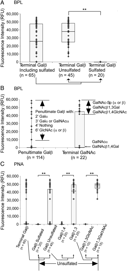
Fig. 4.
Motif comparisons of BPL and PNA. Each point on the scatter plots represents a glycan on the glycan array. The fluorescence after detection with BPL or PNA is plotted for glycans containing particular motifs. (A) The effect of sulfation on BPL binding. The fluorescence is plotted for glycans that contain terminal Galβ and separately for those that are sulfated or unsulfated. The difference between BPL binding to unsulfated and sulfated glycans (indicated by double asterisks) was highly significant (P << 0.001). (B) BPL binding to penultimate Galβ and terminal GalNAc. The scatter plots show the fluorescence signals of all glycans containing either of these two motifs. Additional features of the glycans in each group are indicated. (C) Iterative comparison of motifs bound by PNA. Each group of glycans is a subset of that to the left, beginning with all glycans containing terminal Galβ. PNA showed significantly higher binding to terminal Galβ when the glycan was not sulfated (P = 0.003), followed by significantly higher binding when the glycan was unsulfated and the Galβ was in 1,4 linkage to the penultimate sugar (P < 0.001), followed by significantly higher binding still when the glycan was unsulfated and the Galβ was in 1,4 linkage to a Gal or GalNAc and not a GlcNAc (P << 0.001). All P-values were calculated using the student's t-test.

