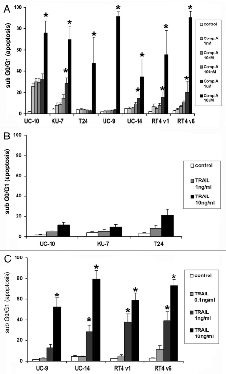
Figure 1.
Flow cytometric analysis of DNA fragmentation by PI staining 24 hours after treatment with compound-A and TRAIL as single agents. (A) Compound-A. No statistically significant difference in apoptosis was seen between control and Compound A at concentrations of 1 nM, 10 nM and 100 nM in all cell lines. At concentrations 1 uM and 10 uM a significant increase in the percentage of apoptotic cells was seen in all cell lines. (*indicates p < 0.05). (B) TRAIL, minimally sensitive cell lines. No statistically significant difference in apoptosis was seen between control and either concentration of TRAIL. (C) TRAIL, highly sensitive cell lines. None of the sensitive cell lines demonstrated a significant increase in apoptosis when exposed to the lowest concentration of TRAIL 0.1 ng/mL (p > 0.05) but statistical significance was achieved for the higher concentrations of TRAIL in every cell line. (p < 0.05) All graphs indicate the Mean ± SE M, three independent experiments.

