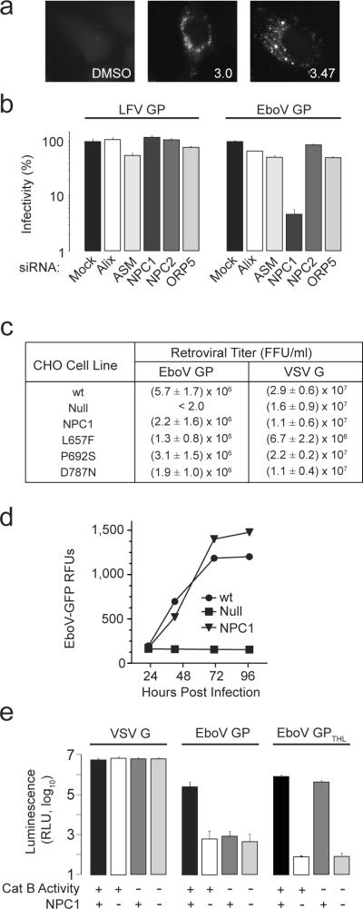
Figure 2. NPC1 is essential for ebolavirus infection.
a, HeLa cells were treated with 3.0 (20 μM), 3.47 (1.25 μM) or vehicle for 18 hours, then fixed and incubated with the cholesterol-avid fluorophore filipin.
b, HeLa cells were transfected with siRNAs targeting ASM, Alix, NPC1, NPC2, and ORP5. After 72 hours, VSV EboV GP or LFV GP infection of these cells was measured as in Fig 1c. Data is mean ± s.d. (n=3) and is representative of 3 experiments.
c, CHOwt, CHOnull and CHOnull cells stably expressing mouse NPC1 (CHONPC1) or NPC1 mutants L657F, P692S, D787N were exposed to MLV particles encoding LacZ and pseudotyped with either EboV GP or VSV G. Results are the mean ± s.d. (n=4) and is representative of 3 experiments.
d, CHOwt, CHOnull, and CHONPC1 cells were infected with replication competent ebolavirus Zaire-Mayinga encoding GFP (moi = 1). Results are mean relative fluorescence units ± s.d. (n=3).
e, CHOwt and CHOnull cells were treated with the cathepsin B inhibitor CA074 (80 μM) or vehicle. These cells were challenged with VSV G particles or VSV EboV GP particles treated with thermolysin (EboV GPTHL) or untreated control (EboV GP). Infection was measured as in Fig 1b. Data is mean ± s.d. (n=9).

