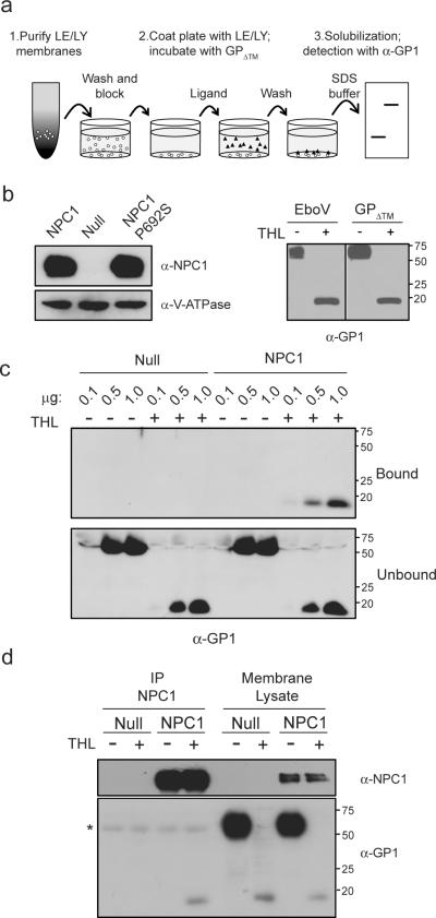
Figure 3. Protease-cleaved EboV GP binds to NPC1.
a, Schematic diagram of EboV GP1 binding assay used in panel c.
b, (left) LE/LY membranes from CHONPC1, CHOnull and CHO NPC1 P692S cells were analyzed by immunoblot using antibodies to NPC1 or V-ATPase B1/2. (right) VSV-EboV GP particles and EboV GPΔ™ protein were incubated in the presence or absence of thermolysin (THL) and analyzed by immunoblot for GP1.
c, EboV GPΔ™ or thermolysin-cleaved EboV GPΔ™ (0.1, 0.5, or 1.0 μg) was added to LE/LY membranes purified from CHOnull or CHONPC1 cells. Membrane bound and unbound GP1 were analyzed by immunoblot.
d,. LE/LY membranes from CHOnull or CHOhNPC1 cells were incubated with EboV GPΔ™ or thermolysin-cleaved EboV GPΔ™. Following binding, membranes were dissolved in CHAPSO, NPC1 was precipitated using an NPC1-specific antibody, and the immunoprecipitate and the input membrane lysate were analyzed by immunoblot for NPC1 (top) or GP1 (bottom). * IgG heavy chain.

