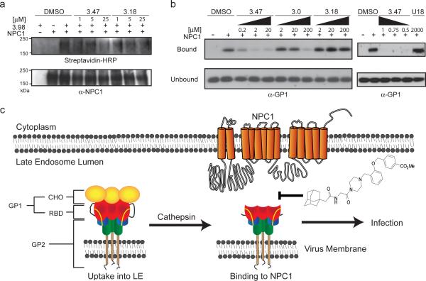
Figure 4. NPC1 is a target of the small molecule inhibitors.
a, LE/LY membranes from CHOnull or CHOhNPC1 cells were incubated at the indicated concentrations of 3.47, 3.18 or DMSO (5%) prior to the addition of the photoactivatable 3.98 (25 μM). After incubation, 3.98 was activated by UV light and then conjugated to biotin. NPC1 was immunoprecipitated and analyzed by immunoblot for conjugation of 3.98 to NPC1 using streptavidin-HRP (top) and recovery of NPC1 (bottom).
b, Thermolysin-cleaved EboV GPΔ™ protein (1 μg) was added to LE/LY membranes from CHOnull or CHONPC1 cells in the presence of DMSO (10%) or the indicated concentrations of 3.47, 3.0, or 3.18 (left panel), and 3.47 or U18666A (U18, right panel). Membrane bound and unbound GP1 were analyzed by immunoblot.
c, Proposed model of EboV entry. Following EboV uptake and trafficking to late endosomes24,25, EboV GP is cleaved by cathepsin protease to remove heavily glycosylated domains (CHO) and expose the putative receptor binding domain (RBD) of GP16,17–19. Binding of cleaved GP1 to NPC1 is necessary for infection and is blocked by the EboV inhibitor 3.47.

