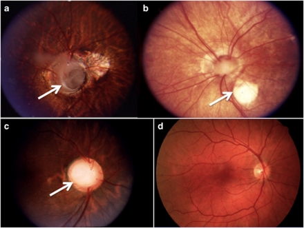
Figure 1.
Fundus photographs from three patients with renal coloboma syndrome (a–c) and a normal retina for comparison (d). (a) Left retina from a patient with RCS and PAX2 mutation c.76dup.14 The arrow denotes a deeply excavated optic disc. (b) Right retina from a patient with RCS and PAX2 mutation c.76delG.14 The arrow denotes a retinal defect, however, the defect is temporal rather than nasal. (c) Right retina from a patient with PAX2 mutation c.76dup.13 The optic disc is enlarged and excavated. In all three retinae from patients with renal coloboma syndrome the retinal vessels emerge from the edge of the disc rather than the center. (d) A normal retina for comparison. Note that the typical optic nerve is smaller, compact and the retinal vessels emerge from the center of the disc.

