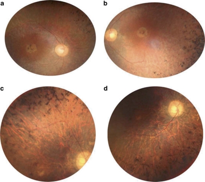
Figure 2.
Fundus images of affected patients in family RP127. (a) Right and (b) left eye posterior poles of patient II.5 at age 42 showing round shape atrophy of the macula, advanced atrophy of the peripheral retina, pigment deposits and narrowing of retinal vessels. (c) Superior and (d), inferior retina in the right eye of patient II.2 at age 54 showing major atrophy of the whole retina including retinal periphery and macula, with pigment deposits, tenuous retinal vessels and waxy pale optic disks.

