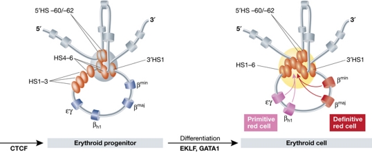
Figure 4.
A schematic representation of the three-dimensional structure of the mouse β-globin locus during differentiation. In erythroid progenitors, a ‘Chromatin Hub’ (CH) is formed by the clustering of the CTCF sites that surround the β-globin locus. Upon differentiation, the remaining hypersensitive sites of the LCR and the globin gene that is activated participate in this clustering to form an ‘Active Chromatin Hub’ (ACH). Hypersensitive sites are represented by the ovals and genes by the cylinders. In grey, the olfactory receptor genes that surround the locus are depicted. A developmental switch in gene usage occurs between primitive and definitive red cells.

