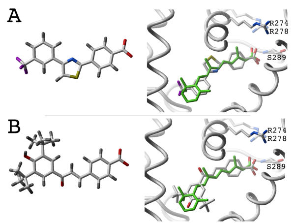
Figure 5.
Structure of the novel RAR agonists. Agonists 1 and 2 are shown (respectively A and B). Left: chemical structure of the compounds. Right: Representation of the compounds docked into the binding pocket of RAR (important residues are displayed as sticks: R274, R278, S289), and superimposed with the crystal structure of all-trans RA (green). The receptor is represented as a white ribbon. Hydrogens are not displayed for clarity. Color coding: carbons, oxygens, nitrogens, sulfurs, fluorides and hydrogens are colored white, red, blue, yellow, magenta and gray respectively.

