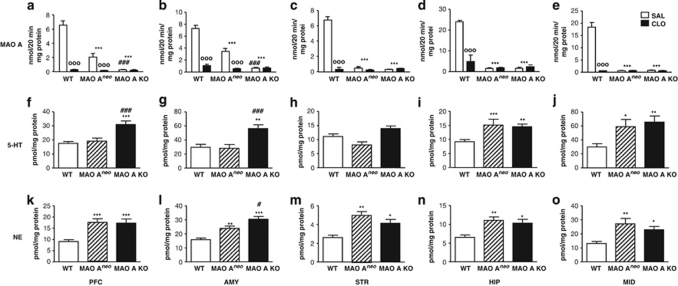
Figure 2.
Brain-regional levels of MAO-A activity, 5-HT and NE in adult WT, MAO-ANeo, and MAO-A KO mice. (a, b) MAO-ANeo mice display higher brain MAO-A activity levels than MAO-A KO mice in prefrontal cortex and amygdala, but lower than WT controls. The low levels of MAO-A were attenuated by teatment with the MAO-A inhibitor clorgyline (10 mg/kg, i.p.) (c–e) Conversely, MAO-ANeo mice showed comparable MAO-A activity in other brain regions tested. (f, g) MAO-ANeo mice exhibit 5-HT levels comparable to WT mice in the prefrontal cortex and amygdala. Although similar 5-HT levels were detected in the (h) striatum in all genotypes, both MAO-A mutants exhibited higher levels of 5-HT in the (i) hippocampus and (j) midbrain. (k–o) NE levels were higher in MAO-ANeo and MAO-A KO mice than in WT mice in all brain regions. MAO-A KO mice display higher NE levels than do MAO-ANeo mice in the amygdala. Values are represented as mean±SEM. *P<0.05, **P<0.01, ***P<0.001 in comparison with WT mice and #P<0.05 and ###P<0.001 in comparison with MAO-ANeo mice. 000P<0.001 compared with saline treatment. AMY, amygdala; CLO, clorgyline; HIP, hippocampus; 5-HT, serotonin; MID, midbrain; NE, norepinephrine; PFC, prefrontal cortex; SAL, saline; STR, striatum.

