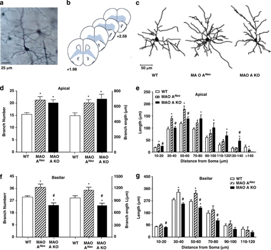
Figure 3.
MAO-ANeo and MAO-A KO mice show a robust increase in dendritic arborization in the orbitofrontal cortex (OFC). (a) Digital light micrograph of Golgi-stained neurons in the OFC of a WT mouse. Scale bar=25 μm. (b) Schematic diagrams of coronal sections through the mouse prefrontal cortex. The coordinates indicate position relative to bregma (Paxinos and Franklin, 2001). Neurons were sampled from the portions of OFC (shaded areas). (c) Computer-assisted reconstructions of Golgi-stained neurons in the OFC of WT, MAO-ANeo, and KO mice. Scale bar=50 μm. (d) The total number and length of apical branches were increased in MAO-ANeo and MAO-A KO mice relative to the WT controls. (e) Both mutant lines showed significant increases in apical dendritic material. (f) Significant increases in basilar branch number and length were detected in MAO-ANeo mice relative to WT, whereas MAO-A KO mice showed decreased number and length of basilar branches. (g) Similarly, MAO-ANeo mice showed increases in basilar dendritic material compared with both WT and MAO-A KO counterparts. The values are represented as the mean±SEM. *P<0.05 in comparison with WT mice; #P<0.05 in comparison with MAO-ANeo mice.

