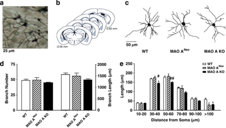
Figure 4.
MAO-ANeo mice show a subtle reduction in dendritic material in the basolateral amygdala (BLA). (a) Computer-assisted reconstructions of Golgi-stained neurons in the BLA of WT, MAO-ANeo, and MAO-A KO mice. Scale bar=25 μm. (b) Schematic diagrams of coronal sections through the mouse amygdala. The coordinates indicate position relative to bregma (Paxinos and Franklin, 2001). Neurons were sampled from the portions of BLA (shaded areas). (c) Computer-assisted reconstructions of Golgi-stained neurons in the OFC of WT, MAO-ANeo, and MAO-A KO mice. Scale bar=50 μm. (d) The total number and length of dendritic branches were not significantly different in MAO-ANeo and MAO-A KO mice relative to their WT counterparts. (e) In MAO-ANeo mice, decreases in dendritic length were subtle and restricted to the most distal branches, whereas MAO-A KO mice showed larger decreases in both proximal and distal dendritic length. The values are represented as the mean±SEM. *P<0.05 compared with WT mice and #P<.05 and ##P<0.01 compared with MAO-ANeo mice.

