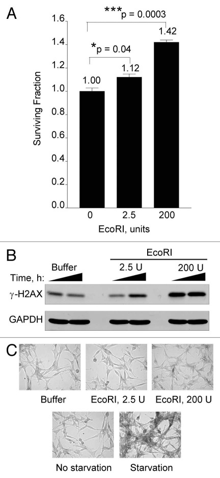
Figure 10.
Low levels of DSBs increase cell proliferation. (A) Human U87 cells were electroporated with buffer alone, 2.5 U or 200 U of EcoRI enzyme as described in the legend to Figure 8. Four days after electroporation cells were collected from triplicate dishes, stained with Trypan Blue and cell viability determined by flow cytometry as described.40 (B) Electroporated U87 cells from (A) were cultured for 1 or 6 h and then collected and processed for western blotting with GAPDH serving as loading control. (C) Electroporated U87 cells from (A) were stained for β-galactosidase (top fields). Starved or unstarved U87 cells were stained for β-galactosidase after 72 h (bottom fields). Data points, survival fraction as a function of electroporated EcoRI. Error bars, SEM; n = 3. Fold (x) denotes changes in live cell levels compared to control (buffer, no EcoRI). *p < 0.05; ***p < 0.0005.

