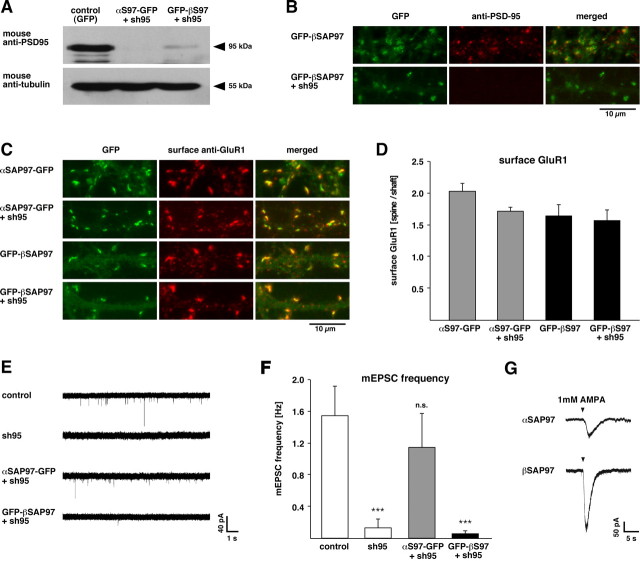
Figure 7.
Isoform-specific rescue of PSD-95 knockdown phenotype. A, Western blot of lysates from dissociated hippocampal neurons infected with lentivirus expressing soluble EGFP for control (left lane), αSAP97–EGFP plus sh95 (middle lane), or EGFP–βSAP97 plus sh95 (right lane) and immunostained with antibodies against PSD-95 (top) or α-tubulin (bottom). Note the dramatic reduction of PSD-95 in neurons expressing the sh95 RNAi. B, Dendrites of neurons infected with EGFP–βSAP97 ± sh95 (green; left) and immunostained with PSD-95 antibodies (red; middle). Merged images (right) show that PSD-95 immunoreactivity colocalizes with βSAP97 in dendritic spines of control neurons (top) but is absent from neurons expressing sh95 (bottom). C, Dendrites of neurons infected with αSAP97–EGFP ± sh95 (top) or EGFP–βSAP97 ± sh95 (bottom) and immunostained with antibodies to label surface GluR1 (red). D, Quantification of surface GluR1 intensity (expressed as spine/shaft ratio ±SEM) in neurons infected with αSAP97–EGFP or EGFP–βSAP97 with or without sh95. Expression of sh95 does not significantly alter surface GluR1 levels in the presence of either SAP97 isoform. E, Sample mEPSC traces from untransfected control neurons (top) and those coexpressing sh95 together with soluble EGFP (sh95), αSAP97–EGFP, or EGFP–βSAP97. F, Quantification of mEPSC frequency data from these cells. The Mann–Whitney Rank sum test determined a significant effect (p < 0.001) of sh95 and EGFP–βSAP97 plus sh95 but not αSAP97–EGFP plus sh95, on mini-EPSC frequency. G, Sample traces of inward AMPAR currents induced by focal AMPA application in α- (top) or βSAP97–EGFP (bottom)-transfected neurons.

