
Figure 8.
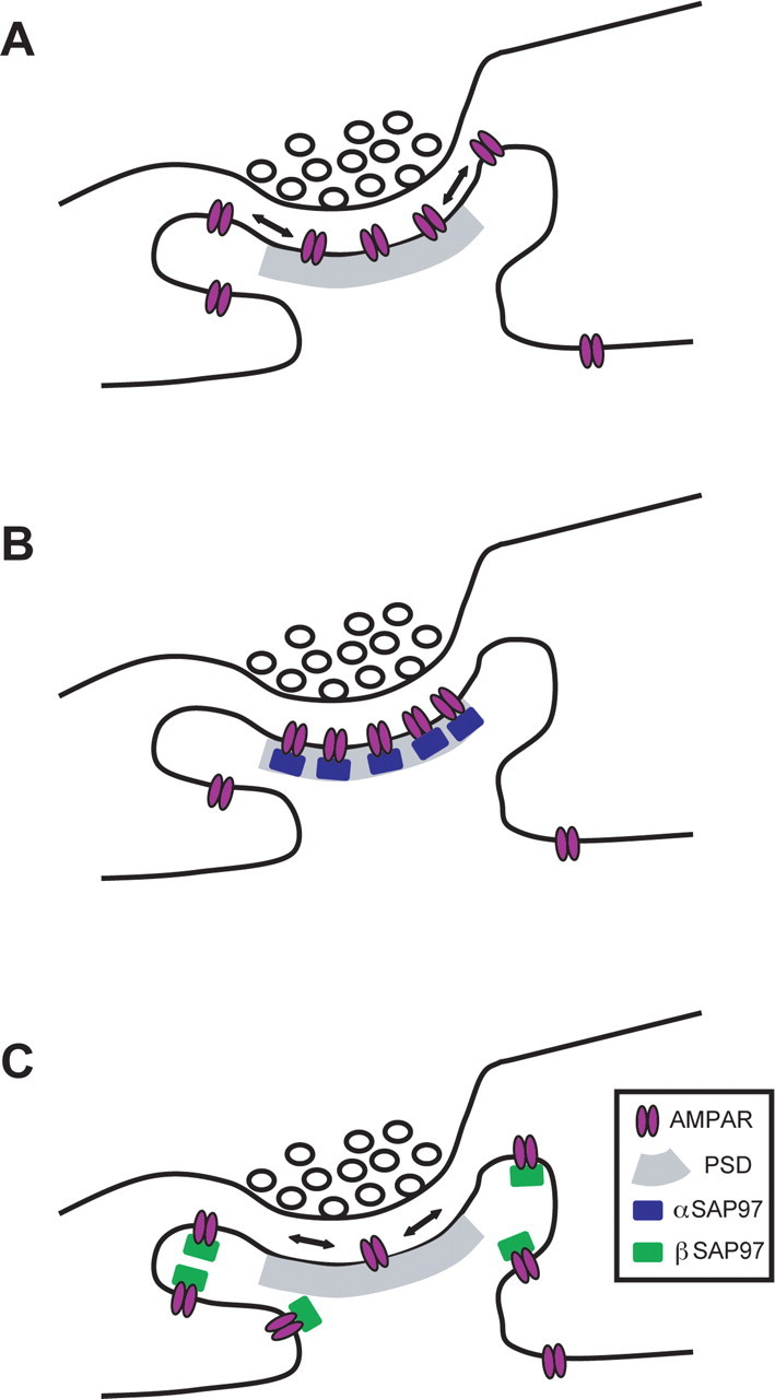
Model of how α- and βSAP97 regulate the subsynaptic localization of GluR1-containing AMPARs. A, Schematic diagram of an excitatory synapse at steady state. Cell-surface AMPARs are concentrated at the PSD through their interactions with PSD-95 but can laterally diffuse to extrasynaptic domains of the spine. B, Excitatory synapse expressing high levels of αSAP97. This isoform localizes to the PSD, creating additional binding sites for surface AMPARs within this domain and restricting their lateral mobility. C, Excitatory synapse expressing high levels of βSAP97. This isoform localizes to extrasynaptic domains of the spine, creating new non-PSD binding sites for surface AMPARs and thereby inhibiting their diffusion into the PSD.