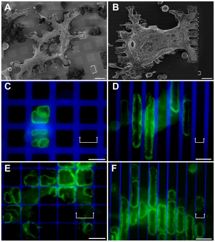
Figure 1. Osteoclasts and sealing zones on vitronectin (VN)/PLL-g-PEG micro-patterns.
(a, b) SEM micrographs of differentiated osteoclasts spreading over non-adhesive PLL-g-PEG areas on square and striped VN-coated micro-patterns: a) 15×15 µm, marked with double arrows, 10 µm-wide barrier; b) 6 µm-wide stripes, marked with double arrows, 1.8 µm-wide barrier, marked by arrowheads). The cells were critical point dried after partial removal of the cell body, to enable better detection of the adhesion pattern on the substrate. (c, e) Osteoclasts growing on 20×20 µm VN patterns separated by (c) 8.5 µm- or (e) 1 µm-wide PLL-g-PEG barriers. Panel c shows a single frame from a time- lapse movie of a GFP-actin expressing osteoclast, while in Panels d-f, actin was stained with phalloidin-FITC. (d, f) Osteoclasts growing on 11 µm-wide VN coated stripes separated by (d) 4.5 µm- or (f) 900 nm-wide PLL-g-PEG barriers. Green: GFP-actin/Phalloidin-FITC, Blue: PLL-g-PEG-TRITC. Scale bars, 20 µm.

