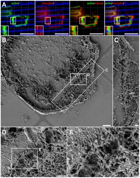
Figure 5. Interconnecting actin fibers bridging over a 1 µm-wide PLL-g-PEG barrier.
(a) SZ formed on striped micro-patterns, bridging a PLL-g-PEG barrier (2.8 µm-wide adhesive stripes, barriers 1 µm wide). Note that only the actin component of the SZ bridges the barrier, while the vinculin domains remain restricted to the adhesive areas. Inserts display magnifications of respective selections. Green: Phalloidin-FITC; Red: anti-vinculin 546; Blue: PLL-g-PEG-Atto-633. Scale bars, 10 µm. (b–e) SEM micrograph of a ventral membrane formed on 40×40 µm VN coated micro-patterns displaying a SZ spanning a 1 µm-wide PLL-g-PEG barrier by interconnecting actin fibers (shown at 4 levels of magnification). (c–e) Magnifications of the respective selections in b and d. Scale bars: (b) 3 µm; (c, d) 1 µm; (e) 200 nm. Images in b, d and e were taken after tilting the stage of the SEM by 30°, to facilitate visualization of the area underneath the fibers.

