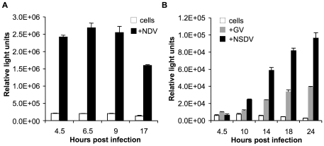
Figure 1. Delayed IFNβ induction in GV and NSDV-infected cells.
Vero cells were transfected with 400 ng of pIFNβ-luc and 200 ng pJATLacZ. After 24 hours of transfection, the cells were infected with (a) NSDV or GV, or (b) NDV at an MOI of 1 TCID50 unit per cell, or left uninfected. At the indicated time points cells were lysed and assayed for luciferase and β-galactosidase activities. The ratio of these two activities was taken as the relative luciferase activity (in RLU). Shown are the data from a representative experiment in triplicates; error bars represent one standard error of the mean.

